ESTABLISHED 1997
ISO9001 CERTIFICATED
Home >> Tungsten Alloy Radiation Shield >> Single Photon Emission Computed Tomography

Single Photon Emission Computed Tomography

Single Photon Emission Computed Tomography

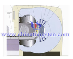
Single photon emission computed tomography (SPECT, or less commonly, SPET) is a nuclear medicine tomographicimaging technique using gamma rays. It is very similar to conventional nuclear medicine planar imaging using a gamma camera. However, it is able to provide true 3D information. This information is typically presented as cross-sectional slices through the patient, but can be freely reformatted or manipulated as required.

The technique requires delivery of a gamma-emitting radioisotope (a radionuclide) into the patient, normally through injection into the bloodstream. A marker radioisotope is attached to a specific ligand to create a radio ligand, whose properties bind it to certain types of tissues. This marriage allows the combination of ligand and radiopharmaceutical to be carried and bound to a place of interest in the body, where the ligand concentration is seen by a gamma camera.
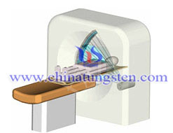
tungsten single photon emission computed tomography tungsten single photon emission computed tomography

The Applications of Tungsten Shielding in Single Photon Emission Computed Tomography

Due to its high density, excellent absorption behaviour against radiation and environmental friendly characteristics, tungsten alloy can be widely used in gamma camera (SPECT) utilize radioactive materials injected as tungsten alloy syringe shield. Radiation is an effective tool within medicine for both diagnostics and treatment of patients. Techniques such as SPECT utilize radioactive materials injected into the patient, which are then monitored by single photon emission computed tomography (SPECT) to detect the presence of tum ours in the body.

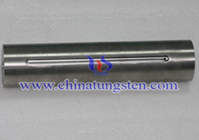
Tungsten radiation shielding is one important component of gamma camera (SPECT) to set radiation resources. Tungsten radiation shielding for SPECT can be used in complement any gamma imaging study, where a true 3D representation can be helpful, e.g., tumor imaging, infection (leukocyte) imaging, thyroid imaging or bone scintigraphy. Because SPECT permits accurate localization in 3D space, it can be used to provide information about localized function in internal organs, such as functional cardiac or brain imaging.

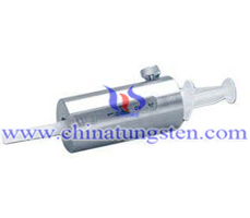
Tungsten radiation shielding also can be used in the syringe shield for SPECT scanner. 2 mm solid tungsten flange helps syringe shield the hand when withdrawing liquid from a vial. Flange is easily removed to allow transition from drawing dose to patient injection. 9 mm thick glass-5.2g/cc gives the greatest protection of any glass in any syringe shield and syringe shield is easily replaced. twist-turn and the syringe is held firmly.

Why Use Tungsten Shielding in Single Photon Emission Computed Tomography?

The advantages of tungsten alloy:
High radiation attenuation; good shielding capability
Thinner and often lighter than equivalent lead shields
Easy to sterilise and keep clean
Non-toxic
Easily machined with conventional tools
Hard and durable – no need for coating

During design of shielding, tungsten alloy radiation shielding is calculated according to requirements of shield to abate the multiple shielding materials' thickness.
Formula: K=e0.693 d / △1/2
K: Shield weakened multiple
△ 1/2: The tungsten alloy radiation shielding material of the half-value layer values
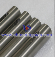
 

tungsten radiation shielding tungsten radiation shielding tungsten radiation shielding

If you have any enquiry or purchase plan about tungsten radiation shielding for single photon emission computed tomography,
please feel free to contact us at sales@chinatungsten.com sales@xiamentungsten.com. Or call: 0086 592 512 9696, 0086 592 512 9595.

Address: 3F, No.25 WH Rd., Xiamen Software Park Ⅱ, FJ 361008,China
Phone:+86-592-5129696,+86-592-5129595; Email:sales@chinatungsten.com sales@xiamentungsten.com
 Certified by MIIT:闽B2-20090025  闽 ICP备06012101号-1
Copyright©1997 - ChinaTungsten Online All Rights Reserved